Flywheel provides customizable, scalable solutions to support multi-modal imaging research in a wide range of therapeutic areas such as CNS, pulmonary, abdominal, and musculoskeletal investigations.
The platform can also accelerate your ophthalmological imaging research through an extensive suite of optical coherence tomography (OCT) annotation tools and customization options.
Multiple OCT File Type Support
As part of our commitment to provide maximum flexibility and customization options to researchers, Flywheel’s OCT suite of tools simplifies the capture of raw OCT images that come from multiple equipment manufacturers, and seamlessly converts them into more publicly viewable and accessible formats, including DICOM.
This feature can be set up to run automatically on ingest, and the file image types supported by Flywheel’s OCT annotation tools include .png, .jpeg, and .mp4.
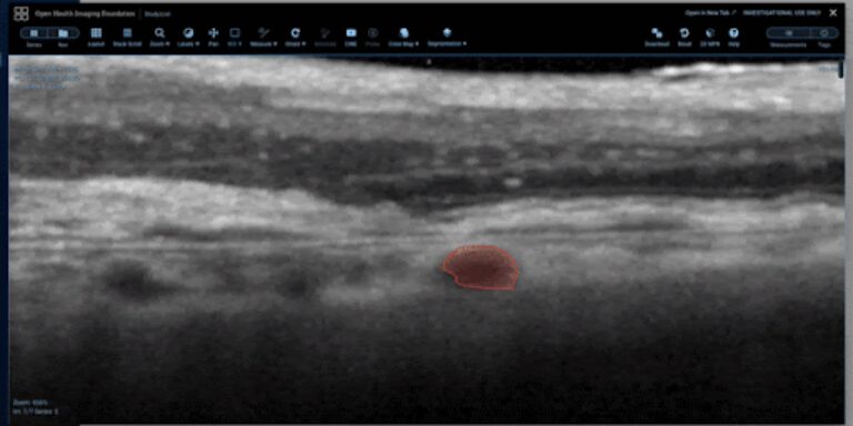
View and Annotate OCT Images
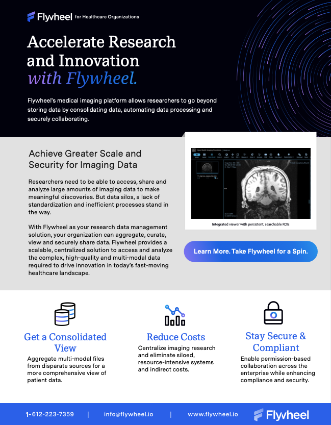
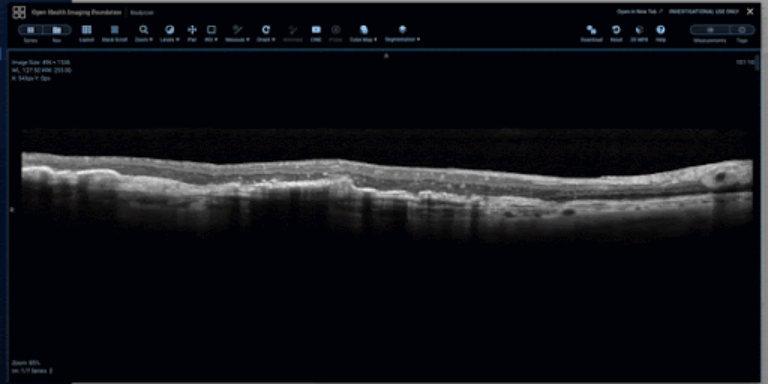
Flywheel offers an integrated, radiology-grade viewer with several powerful features, such as drawing persistent, searchable regions of interest (ROIs) on OCT images, with intuitive and customizable annotations.
As shown below, the viewer supports an open freehand tool for retinal layer tracing, a closed freehand ROI tool for capturing distinct findings, such as intraretinal fluid pockets, and a sculpt tool for precise editing of freehand ROIs.
Open Freehand ROI tool for retinal layer tracing.

Closed Freehand ROI tool for capturing distinct findings.
Sculpt tool for precise editing of freehand ROIs.
Additionally, Flywheel allows customizable asymmetric hanging protocols, such as the ability to have 2D retinal layer imaging and 2D multiplanar reconstruction (MPR) views in the same hanging protocol, permitting linked navigation between 2D and 2D MPR.
Flywheel’s suite of OCT tools also supports slice-specific workflows, where, for example, a study PI can choose to have image readers annotate every third slice. By doing so, other slices are set to “read only” so readers are unable to draw ROIs and no forms would display for them to complete. Once they navigate to a slice that has been promoted for specific workflows, it becomes “read-write” and a special form displays for that single slice.
The viewer’s toolbar configuration can be customized to either include or exclude any of the tool options using “except” or “only” logic. For example, selecting Measurements and specifying “only” and “length” would remove angle and bidirectional tools from the menu. Fully customizable “hot keys” allow you to increase your efficiency by defining keystrokes and mouse actions for tools such as WWWC presets and UI actions.
Support for Data Quality and AI Research
Flywheel’s imaging research solutions can be easily used for AI research frameworks, allowing imaging researchers to construct configurable workflows for multiple readers, while simplifying measurements, data capture, and workflow status.
The OCT viewer supports blinded reader studies with customizable labels and annotations and task assignment workflows. You can customize forms in the viewer for clinical data capture. Customized workflow configuration forms can include data capture using logic rules such as required answer (annotations, measurement, etc.), multiple choice, and conditional display of a field based on a previous answer.
Administrators can configure Flywheel’s platform to automatically assign readers tasks via Gears. The collected form data and annotation is automatically saved and indexed on the session, and can be collected back to master projects. This allows you to perform a search, find other sessions with similar pathologies and enable re-use of data sets for future studies to create training and learning data sets for AI and machine learning applications.
Request a Demo
Flywheel’s OCT viewing solutions have the robust features, powerful tools, and customization options to accelerate multi-modal, multi-reader imaging research in ophthalmology, ocular oncology, oculoplastics, and other fields undertaking large-scale ophthalmological imaging research.
Request a demo to learn how Flywheel can accelerate your ophthalmological research.